Coulomb attraction driven spontaneous molecule-hotspot pairing

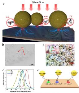
Figure 1. Basic structure and working mechanism of SM-SERS substrate. a) Schematic diagram of rapid SM-SERS signal formation. b) SEM image of SM-SERS substrate. c) Optical image of sample under micro-Raman spectrometer.
Enabling universal, fast, and large-scale uniform single-molecule Raman spectroscopy.
CHENGDU, SICHUAN, CHINA, August 4, 2025 /EINPresswire.com/ -- Raman spectroscopy is based on the principle of inelastic light scattering, enabling precise analysis and identification of materials by detecting spectral characteristics of low-frequency modes such as vibrations and rotations in molecules, solids, and two-dimensional materials. This technology offers advantages including label-free detection, rich information content, and non-destructive analysis, demonstrating broad application prospects in chemistry, biology, medicine, environmental science, and other fields. However, as a higher-order light-matter interaction process, Raman scattering has an inherently tiny scattering cross-section, with signal intensity far lower than fluorescence and Rayleigh scattering. This fundamental limitation severely constrains the detection sensitivity. Developing single-molecule Raman spectroscopy (SM-RS) to achieve Raman signal intensity comparable to fluorescence signals has become an important goal in this field. SM-RS not only represents an ultra-high sensitivity version of traditional Raman spectroscopy but also provides a unique window for observing subtle spectral phenomena in individual molecules. By avoiding interference from statistical averaging effects, this technology can reveal dynamic behaviors and spectral characteristics at the single-molecule level, providing unprecedented precision and depth for molecular science research.The development of surface-enhanced Raman spectroscopy (SERS) has provided an important pathway for achieving single-molecule Raman detection. Among these approaches, electromagnetic enhancement (EME) utilizes localized surface plasmon resonance phenomena in metallic nanostructures to generate strong local electric field enhancement in nanoscale “hotspot” regions, achieving signal amplification of up to 10-11 orders of magnitude. Chemical enhancement effect (CME) significantly increases the Raman scattering cross-section of molecules through charge transfer and chemical bonding interactions between molecules and substrates. Although each of these mechanisms can provide significant signal enhancement individually, a single mechanism typically cannot achieve the enormous enhancement factor of 14-15 orders of magnitude required for ideal single-molecule detection. Fortunately, when two or more of these strategies work together synergistically and constructively, the situation becomes very encouraging.
The research team led by Professor Zhi-Yuan Li at South China University of Technology previously constructed a WS₂-Au nanogap system and discovered that EME from plasmonic hotspots can reach 9-11 orders of magnitude, while two-dimensional (2D) material WS₂ monolayers provide additional CME of 4-5 orders of magnitude. The synergistic effect of EME and CME established an important foundation for achieving single-molecule Raman spectroscopy detection [PhotoniX 5, 3 (2024)]. However, the core scientific questions regarding the mechanism, applicability, detection stability, and enhancement uniformity of this EME-CME synergy enabled SM-SERS still requires in-depth exploration.
Addressing these key issues, Prof. Li and his team propose in this work a Coulomb attraction-driven spontaneous “molecule-hotspot” pairing mechanism, which is the core element for realizing SM-SERS schemes, enabling efficient synergistic action between the enormous CME of WS2 2D materials and the enormous EME of plasmonic nanogaps. Furthermore, utilizing near-infrared laser (785 nm) excitation for Raman signals can effectively suppress fluorescence background interference. The SM-SERS substrate consists of three key components (Figure 1a): gold nanosphere particles (Figure 1b) positioned on a gold thin film mirror forms plasmonic nanogaps, with a 2 nm thick SiO2 separation layer in between to optimize EME; 2D monolayer WS2 single crystal flakes (Figure 1c) sandwiched between SiO2 and gold nanoparticles providing CME; Raman analyte molecules tightly bound to WS2 flakes while simultaneously located in plasmonic hotspots, achieving synergistic EME and CME.
Highlights from the article:
(1) Coulomb Attraction-Driven Spontaneous “Molecule-Hotspot” Pairing
This study first confirms the charge Coulomb attraction-driven mechanism of spontaneous molecule-hotspot pairing through Zeta potential measurements (Figure 1d). Experiments reveal that RhB, R6G, and CV molecules are positively charged while gold nanoparticles are negatively charged. The Coulomb attraction between them drives gold nanoparticles to precisely position directly above molecules already bound to WS2 single crystal flakes, spontaneously forming optimal plasmonic nanogap configurations (Figure 1e). This charge Coulomb force-driven self-assembly mechanism provides a crucial physical foundation for EME-CME synergy enabled SM-SERS, greatly improving the universality, uniformity, robustness, and stability of synergistic SM-SERS active sites.
(2) Near-Infrared Laser Effectively Suppresses Fluorescence Interference, Significantly Enhancing Detection Performance
The study employs 785 nm near-infrared laser as the excitation source, cleverly avoiding strong fluorescence background interference generated by RhB, R6G, and CV molecules under visible light excitation. Compared to 532 nm laser excitation, 785 nm excitation not only eliminates fluorescence baseline contamination but also increases Raman signal intensity by approximately 100-fold, with significantly improved signal-to-noise ratio (Figure 2). This fluorescence suppression strategy, combined with the synergistic effect of electromagnetic and chemical enhancement, provides crucial technical support for achieving ultra-low concentration single-molecule detection, becoming an important complementary pathway for advancing SERS toward the fundamental SM-SERS goal.
(3) Large-Area Uniformly Distributed Rapid Single-Molecule Detection Capability
Through systematic Raman mapping studies on 5 mm × 5 mm large-area substrates, the uniform distribution characteristics of SM-SERS active sites at macroscopic scales are verified. At extremely low concentrations of 10-16 M, 3-4 Raman active sites can be stably observed within each 60 μm × 90 μm micro-region, increasing to 10-20 active sites at 10-14 M concentration, demonstrating good concentration response characteristics. Through systematic studies of different concentration gradients (10-16 M to 10-12 M) (Figure 3), it is found that within the single-molecule detection range, all Raman active sites exhibit similar signal intensity levels, confirming true single-molecule detection capability. This scheme successfully achieves universal detection of three typical Raman molecules: Rhodamine B (RhB), Rhodamine 6G (R6G), and Crystal Violet (CV), with outstanding advantages including low detection sensitivity (10-16 M), fast detection speed (50 ms), good stability, and large-area uniformity, providing important support for practical applications of this SM-SERS technology.
This work is published in Opto-Electronic Advances 2025, Issue 7, with title “Coulomb attraction driven spontaneous molecule-hotspot paring enables universal, fast, and large-scale uniform single-molecule Raman spectroscopy”.
About the Author:
Zhi-Yuan Li, corresponding author of this paper, China National Natural Science Foundation for Distinguished Young Scholars recipient, Professor and Vice Dean of the School of Physics and Optoelectronics at South China University of Technology, Director of the Artificial Light and Sound Microstructure Physics Laboratory. He has been engaged in theoretical, experimental, and applied research in micro-nano photonics, nonlinear optics, laser technology, topological photonics, and quantum physics for nearly 30 years. He has published nearly 530 SCI papers in physics, optics, chemistry, and materials science journals including PRL, Sci. Adv., Nat. Commun., Chem. Soc. Rev., JACS, Adv. Mater., Nano Lett., ACS Nano, Light, OEA, PhotoniX, Adv. Photon., with over 34,000 SCI citations and an H-index of 90.
Read the full article here: https://www.oejournal.org/oea/article/doi/10.29026/oea.2025.240309
Andrew Smith
Charlesworth
+44 7753 374162
marketing@charlesworth-group.com
Visit us on social media:
LinkedIn
YouTube
Other
Legal Disclaimer:
EIN Presswire provides this news content "as is" without warranty of any kind. We do not accept any responsibility or liability for the accuracy, content, images, videos, licenses, completeness, legality, or reliability of the information contained in this article. If you have any complaints or copyright issues related to this article, kindly contact the author above.


